Understanding Pulmonary Interstitial Edema on Chest X‑Ray
When a clinician orders a chest X‑ray, the goal is often to spot fluid where it doesn’t belong. Pulmonary interstitial edema is one such finding—fluid that accumulates in the lung’s supporting tissue rather than the air‑filled alveoli. Recognizing its subtle clues on an X‑ray can steer treatment toward the heart, kidneys, or other underlying problems before they spiral.
What Is Pulmonary Interstitial Edema?
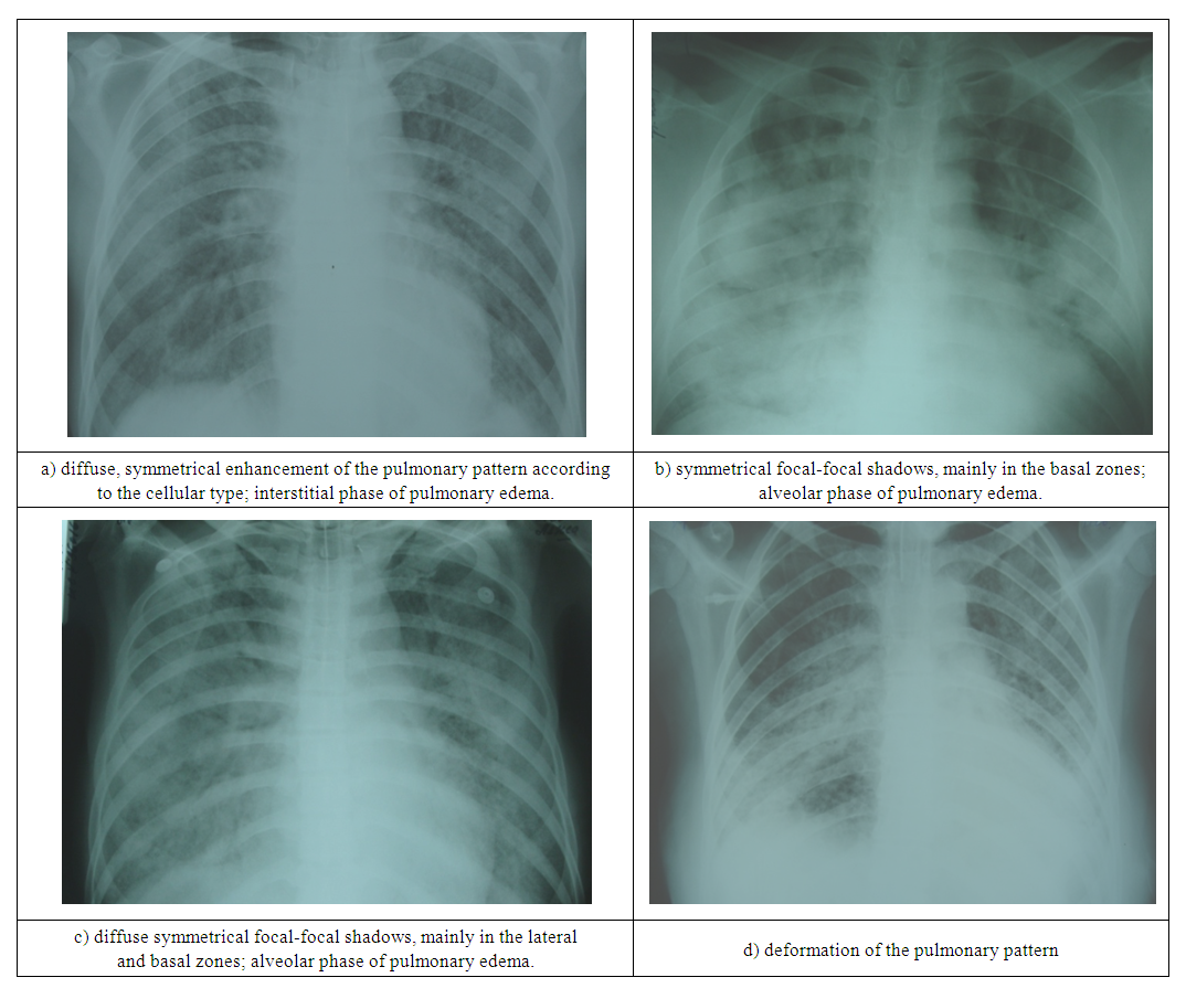
In simple terms, interstitial edema means fluid has leaked into the connective tissue that cushions the tiny air sacs and blood vessels. Unlike alveolar edema, which fills the air spaces and produces a classic “wet” appearance, interstitial fluid stays in the scaffolding, making the radiographic signs more nuanced. The condition often signals the early stages of heart failure, but it can also arise from severe infections, high‑altitude exposure, or inflammatory lung diseases.
How Pulmonary Interstitial Edema Looks on an X‑Ray
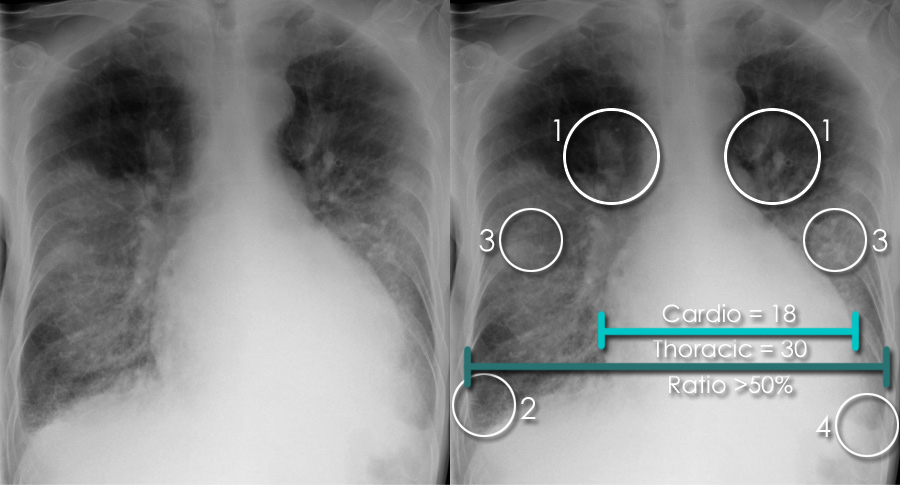
The chest X‑ray is a two‑dimensional snapshot, so the patterns we rely on are indirect. Radiologists look for a handful of tell‑tale signs that together suggest fluid in the interstitium:
- Kerley B lines – thin, horizontal lines at the lung bases, usually 1 cm long, representing thickened interlobular septa.
- Perihilar haze – a hazy increase in density around the central lung zones, often described as “bat‑wing” or “butterfly” pattern when bilateral.
- Reduced vascular markings – the peripheral lung fields may appear less distinct because fluid blurs the normally sharp edges.
- Cardiomegaly – an enlarged heart silhouette frequently accompanies interstitial edema, hinting at a cardiac origin.
These features are rarely seen in isolation; a combination strengthens the suspicion. The findings may be subtle on a first‑time X‑ray, which is why a repeat or a lateral view can sometimes reveal what a single frontal image misses.
Why Early Detection Matters
Spotting interstitial edema early can change the clinical trajectory. Fluid in the interstitium often precedes full‑blown pulmonary congestion, giving physicians a window to adjust diuretics, manage blood pressure, or address an inciting infection. Delayed recognition, on the other hand, may allow the edema to progress into alveolar flooding, where patients develop severe shortness of breath and require intensive respiratory support.
Common Causes and Risk Factors
Not every case of interstitial fluid points to heart failure. Here are the most frequent culprits:
- Left‑sided heart failure – increased hydrostatic pressure pushes fluid outward into the lung’s interstitium.
- Renal dysfunction – when kidneys can’t excrete excess fluid, it accumulates systemically, including in the lungs.
- Acute respiratory distress syndrome (ARDS) – inflammatory leakage can manifest first as interstitial edema.
- High‑altitude exposure – reduced atmospheric pressure encourages fluid shift into lung tissue.
- Inhalation injuries or severe pneumonia – inflammation can compromise the capillary barrier.
Patients with hypertension, diabetes, or a history of coronary artery disease are especially prone, as these conditions often coexist with cardiac or renal insufficiency.
When to Seek Further Evaluation
An X‑ray suggesting interstitial edema should trigger a broader work‑up. Typical next steps include:
- Blood tests for brain‑natriuretic peptide (BNP) or NT‑proBNP to gauge cardiac strain.
- Echocardiography to assess ventricular function and valve integrity.
- Renal panel to check for fluid overload from kidney impairment.
- Possibly a high‑resolution CT scan if the X‑ray findings are equivocal or if a specific interstitial lung disease is suspected.
In the emergency setting, oxygen saturation, respiratory rate, and blood pressure help determine whether immediate intervention—such as intravenous diuretics or non‑invasive ventilation—is warranted.
Practical Tips for Interpreting the Film
Even non‑radiologists can improve their read of a chest X‑ray with a few habits:
- Start by confirming proper positioning—rotation or under‑exposure can mask subtle lines.
- Scan from the apex to the diaphragm, noting any linear opacities that run parallel to the pleura.
- Compare the right and left lungs; asymmetry often points to a focal process rather than generalized edema.
- Remember that Kerley B lines are most visible in the lower zones; if you see them higher up, consider other interstitial diseases.
Practice, coupled with a systematic approach, gradually builds confidence in catching these early signs.
Frequently Asked Questions
Can a normal chest X‑ray rule out interstitial edema?
Not entirely. Early interstitial changes can be so faint that they evade detection on a standard film. A high‑resolution CT or a repeat X‑ray after a few hours may reveal progression that was initially invisible.
Is pulmonary interstitial edema always caused by heart failure?
No. While left‑sided heart failure is the leading cause, renal disease, ARDS, high altitude, and certain infections can also produce interstitial fluid accumulation.
Do Kerley B lines disappear after treatment?
Often they do, especially when the underlying cause—like fluid overload—is corrected. However, chronic interstitial lung disease can leave residual lines even after the edema resolves.
Should I be worried if I see mild interstitial changes on my X‑ray?
Mild findings merit follow‑up. Your doctor will likely order labs or an echo to pinpoint the cause. Early intervention can prevent worsening symptoms and reduce hospital stays.