Understanding G.V. Black’s Cavity Classification: A Deep Dive
When you hear the name G.V. Black, most dental professionals think of the “father of operative dentistry.” His classification system for dental cavities—often called G.V. Black’s cavity classification—remains a cornerstone of both teaching and clinical decision‑making. Though the system was devised over a century ago, it still frames how we describe lesion location, choose restorative materials, and plan treatment. Below, we unpack each class, highlight the nuances that separate them, and explore why the classification continues to shape modern practice.
What Is G.V. Black’s Cavity Classification?
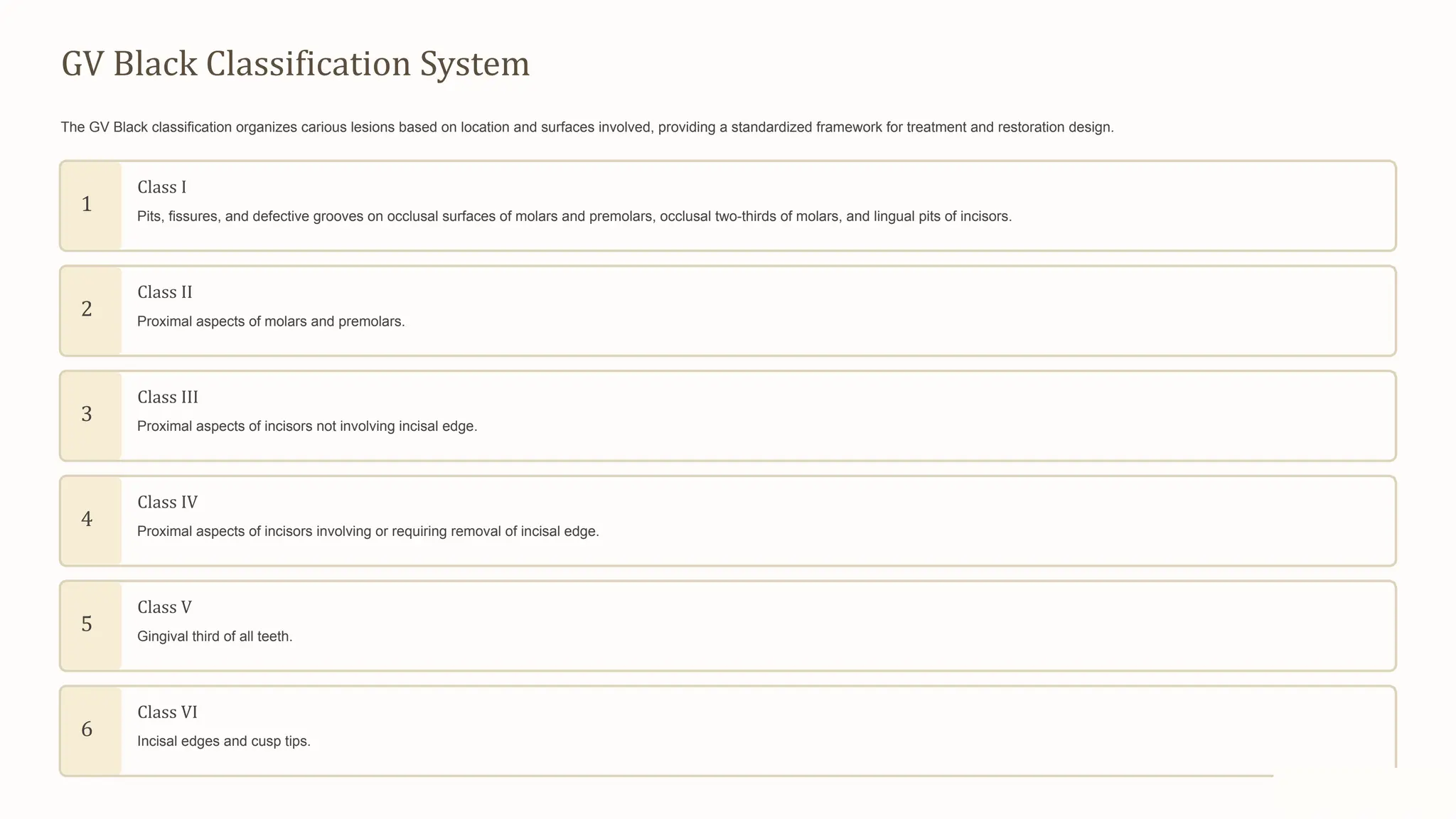
Developed in the early 1900s, Black’s scheme groups carious lesions into six distinct categories based on the tooth surface involved and the extent of tissue loss. The primary goal was to provide a common language that could guide cavity preparation, restoration design, and ultimately, predict long‑term outcomes. While newer diagnostic tools (like digital radiography and laser fluorescence) have added layers of detail, the six‑class framework remains a quick reference point in every dental chart.
Class I: Pit and Fissure Caries
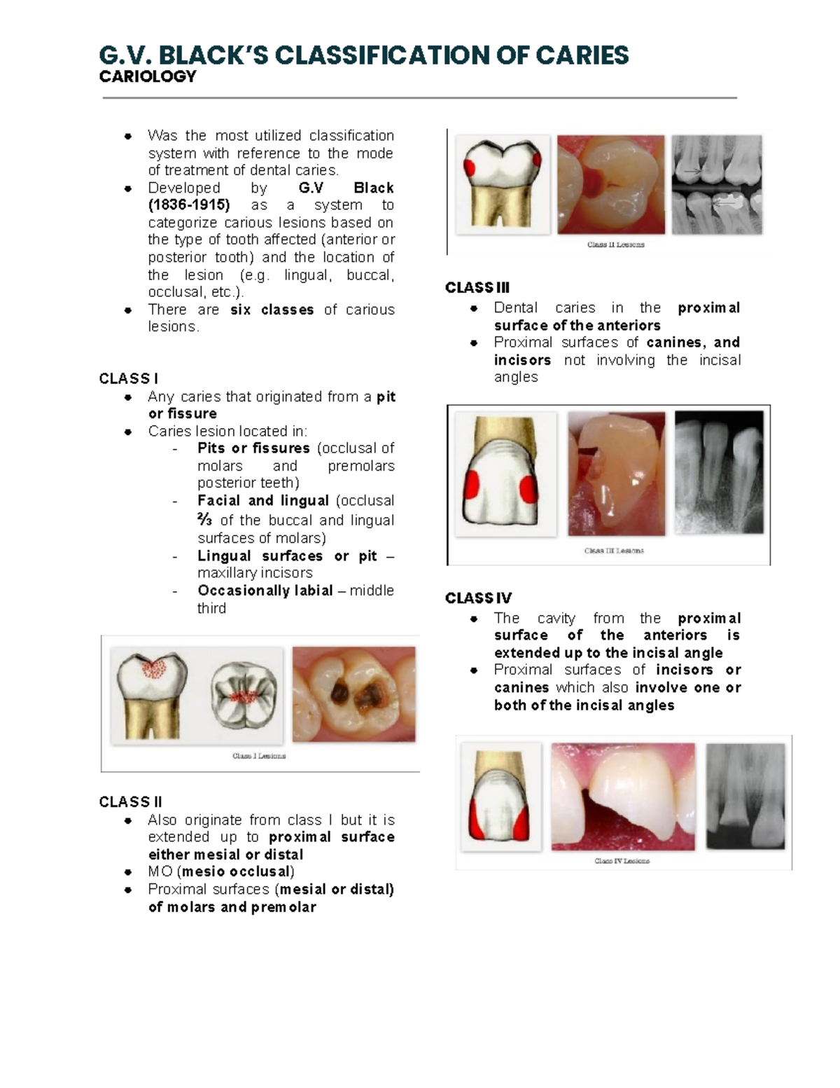
Class I lesions affect the occlusal (chewing) surfaces of posterior teeth, the buccal pits of molars, and the lingual pits of maxillary incisors. These areas are riddled with natural grooves that trap plaque and make thorough cleaning difficult. When decay progresses, it typically starts at the enamel‑dentin junction and may require a simple occlusal filling.
- Typical sites: Molars and premolars’ chewing surfaces, mandibular incisor lingual pits.
- Common restorative choice: Composite resin for aesthetic demands, amalgam where cost is a concern.
- Key challenge: Achieving adequate isolation to prevent moisture contamination during bonding.
Class II: Proximal Caries
Class II cavities involve the mesial or distal surfaces of posterior teeth, extending from the enamel into the dentin and often reaching the occlusal groove. Because these lesions lie between teeth, they are less visible and harder to detect without radiographs. Restorations usually require a box preparation that restores the contact point and maintains proper occlusion.
- Typical sites: Mesial or distal surfaces of molars and premolars.
- Restorative options: Direct composite, indirect onlay, or gold in high‑stress situations.
- Consideration: Ensuring the new restoration re‑establishes a tight interdental contact to prevent food impaction.
Class III & IV: Cervical and Anterior Lesions
Class III lesions affect the interproximal area of anterior teeth without involving the incisal edge, while Class IV lesions extend to include the incisal edge. Both types demand a balance between strength and aesthetics. The decision to place a simple filling versus a more extensive veneer often hinges on the size of the lesion and the patient’s cosmetic expectations.
- Class III: Small to moderate lesions confined to the labial or lingual interproximal area.
- Class IV: Larger lesions that reach the incisal edge, sometimes requiring a buildup of tooth structure.
- Materials: Nanohybrid composites for most cases; ceramic or porcelain for extensive structural loss.
Class V: Root Surface Caries
Class V cavities appear on the gingival third of the facial or lingual surfaces of any tooth, typically where the cementum meets the enamel. These lesions are frequently linked to recession or poor oral hygiene and can be sensitive to temperature changes. Because the area is close to the gingiva, moisture control and soft‑tissue management become critical.
- Risk factors: Gum recession, aggressive brushing, and xerostomia.
- Restorative approach: Light‑cured composite with a bonded technique to seal the margin and protect the root surface.
- Longevity tip: Use a fluoride‑releasing material when the patient has a high caries risk.
Why the System Still Matters Today
Even with digital imaging, 3‑D printing, and minimally invasive techniques, Black’s classification offers a quick, universally understood shorthand. It helps clinicians communicate with dental labs, insurance providers, and patients alike. Moreover, the framework aligns neatly with contemporary concepts such as minimally invasive dentistry, where the goal is to preserve as much healthy structure as possible while still addressing the lesion effectively.
Modern adaptations often add sub‑categories—like “Class VI” for developmental defects or “Class VII” for hypoplastic pits—but the original six classes remain the backbone of most dental curricula. By mastering the fundamentals, a practitioner can more readily incorporate newer technologies without losing the clarity that Black’s system provides.
Frequently Asked Questions
Q: Are Black’s classes still taught in dental schools?
A: Yes. All accredited programs include the six‑class system in operative dentistry courses, using it as a foundation before introducing digital diagnostic tools.
Q: How does the classification affect insurance coding?
A: Many dental insurance forms require the cavity class to determine coverage levels, especially for restorative versus cosmetic procedures.
Q: Can a single tooth have lesions from multiple classes?
A: Absolutely. It’s common for a molar to exhibit a Class I pit caries on the occlusal surface and a separate Class II proximal lesion, each needing its own treatment plan.
Q: Does the classification guide material selection?
A: While not prescriptive, the class hints at functional demands—e.g., high‑stress posterior sites (Class II) often benefit from stronger composites or indirect restorations.